On this interview, Information-Medical talks to a pioneer of glycan imaging, Richard Drake about glycans, their position in sustaining mobile homeostasis, glycopeptide evaluation, and technique growth for glycan imaging.
What are glycans, and why are they essential to biology?
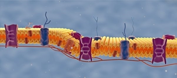
Glycans are advanced carbohydrates derived from fundamental glucose and glutamine metabolism. These glycans are metabolized in advanced carbohydrate constructions on the cell floor, creating an outer shell on the cell floor termed the ‘glycocalyx.’
Functionally, glycans are concerned in protein folding, protein sorting, protein secretion, cell-cell recognition on the floor, organelle and mobile localization, and, extra importantly, immune recognition.
What’s there to be taught from mapping glycan distributions in tissues?
Glycans on the cell floor assist to take care of regular mobile homeostasis and well being generally. In the event that they get modified, particularly in illness eventualities, they have an inclination to get a lot bigger and complicated.
These are splendid targets for MALDI imaging, particularly in case you have the tissues for illness states. This additionally hyperlinks effectively with rising single-cell strategies to take a look at glycan profiles and medical imaging and radiology.
Monitoring Tumor Development by Glycan Imaging Mass Spectrometry – Prof. Richard Drake
Monitoring Tumor Development by Glycan Imaging Mass Spectrometry – Prof. Richard Drake from AZoNetwork on Vimeo.
Does glycan imaging have any clear benefits over different molecular imaging strategies?
There are various sensible benefits to glycan imaging; its easy, quick, reproducible and strong sign detection is at its core.
One of many benefits of the assay is that we’re spraying an enzyme that creates the sign we detect, so we solely detect what the enzyme cleaved. We’re additionally discovering that there are 25 to 30 glycans which are very robustly detected in something that might be thought of alive. This technique could be very reproducible and really definable.
These glycans can differ of their intensities, and that is the place we get into illness variations. This permits us to work with bigger medical cohorts, making our research reproducible and comparatively quick. This opens up loads of medical diagnostic alternatives.
The assay works on formalin mounted tissues in order that we are able to get these immediately from pathology collections. Tens of millions of those tissues can be found, so discovering clinically essential tissues to run has at all times been easy.
Picture Credit score: Marco G Faria/Shutterstock.com
Have you ever discovered any correlations between particular person glycans and mobile illnesses? In that case, would you describe it as being extra diagnostic or prognostic?
We’re discovering that they’re each probably diagnostic, in addition to prognostic. There may be growing proof that the degrees of ‘excessive mannose constructions’, that are biosynthetic precursors to all the opposite advanced glycans, are indicative of the metabolism of a tumor cell or an immune cell, as an illustration.
We’ve additionally seen that particularly tumor subtypes – for instance, a mucinous or a neuroendocrine tumor – there are very particular lessons of glycan constructions. The constant hallmark of those constructions are the presence within the tumor of enormous tetraantennary N-glycans with a number of fucose sugars. Apparently, the numbers of fucose residues varies with organ kind, in addition to different sugar modifications.
What are you able to say in regards to the correlation between distributions of glycans and originating glycoproteins?
We’re discovering that there are various correlations between biofluids and tissues, and defining these is the topic of ongoing analysis.
We discovered that the strategy works so effectively on tissues that we may export it to biofluids, which has expanded into many alternative applied sciences. We’ve a technique now the place we are able to profile nearly any biofluid for glycan content material.
As well as, we discovered that we may adapt this technique to antibody arrays. This opened up an entire new avenue the place we may goal serum glycoproteins and glycotype, for instance, for every serum glycoprotein. This was by no means doable in an array-type format.
Now that we now have the power to take a look at the biofluids, whether or not by way of complete glycan profiling or with the antibody arrays, we shaped GlycoPath as a method and car to develop medical diagnostics on the biofluid degree; but in addition to hyperlink that again to tissues.
As we use tissues which have come straight from pathology, we’re more and more utilizing immunohistochemistry strategies to hyperlink the glycans with a selected pressure of a glycoprotein or goal.
Extra lately, we now have been taking a look at COVID-infected tissues, and we now have been capable of hyperlink the place the spike glycoprotein and immune cell clusters are with completely different glycans which are current.
We’ve additionally began taking a look at basic tumor antigens which are carbohydrate-based in pathology and immunohistochemistry phrases. We have been capable of hyperlink these with tumors and the glycans that we detected.

Picture Credit score: Alpha Tauri 3D Graphics/Shutterstock.com
To researchers finding out protein distributions utilizing frequent tissue imaging methods like immuno-based imaging, would glycan imaging give elevated perception into illness pathways?
Completely. In the meanwhile, I can not consider any state of affairs the place glycan imaging wouldn’t complement present immunostaining strategies. Signaling regulators, oncogenes, development issue genes, transcripts or, certainly, something that’s altering the exercise of the cell goes to point out up within the glycan code on the floor.
This technique is especially helpful given the CRISPR-Cas9 revolution happening with all of the genetic fashions that may be made by knocking in or knocking out particular person genes.
In case you take any CRISPR-Cas9 animal and knock in or knock out a gene concerned in metabolism or development regulation, a change in glycan shall be noticed.
Most, if not all, present most cancers immunotherapies are focusing on floor glycoproteins that regulate immune operate. All of those immune checkpoint proteins are glycosylated, and it’s already documented that adjustments within the glycosylation of those targets additionally have an effect on the remedy final result.
Going after these immunotherapy-treated tissues is certainly one of our main targets shifting ahead.
From all of the glycan pictures produced in your lab, what remark would you say has been most fun?
Asking which remark is my favourite is like asking what my favourite beer is – it’s a very time and place dependent reply, and the default response is ‘the beer I haven’t had but.’ It’s the identical with glycan imaging. Each time I run a brand new tissue, there’s something new and thrilling within the outcome.
Nevertheless, there have been some milestones, significantly in 2012 once we did the primary picture that confirmed that the strategy was working, and in 2013, once we ran the primary FFPE tissue and confirmed it may work with archived tissues. These moments opened up the floodgates for lots of different strategies.
One other spotlight was the primary time we ran a neuroendocrine tissue from the prostate. I may inform instantly that this was very completely different from all the things else we had analyzed thus far. It was the primary time we had seen the big numbers of tumor related glycans with a number of fucose residues. We now know it is a hallmark of neuroendocrine tumors from all tissue varieties.
It has additionally been nice to work with Joe Ippolito, a radiologist at Washington College, who offered the neuroendocrine tumor tissues. This collaboration has morphed into many research the place we try to hyperlink what we’re seeing in all these prostate tumors with medical imaging.
Going within the different course in the direction of single-cell evaluation, we’re performing some very thrilling work with Mike Angelo at Stanford College, linking the glycan imaging along with his multiplexed cell and immune focused probes immediately on the identical tissues. That is opening up an entire new avenue for multimodal evaluation.
Just lately, we now have simply began a collaboration with nephropathologists Jan Braesen and Jessica Schmitz at Hanover Medical College, and so they offered some incredible new tissues within the immunotherapy area. I’m very excited in regards to the preliminary outcomes and the place that is going.
As a pioneer of glycan imaging, what have you ever discovered to be the most important false impression that stops researchers from beginning a glycan imaging examine?
I believe the most important false impression about beginning glycan imaging begins with one phrase: glycophobia. Worry of the structural and organic complexity and nomenclature related to glycans. There are loads of causes for this challenge – a few of these causes are academic as a result of I don’t suppose that is particularly taught anymore at most establishments.
Even for those who begin studying about this discipline, there are loads of archaic phrases and an entire different language and vocabulary to cope with. It appears sophisticated as a result of it’s not a template-driven synthesis.
To assist folks with their glycophobia, we conduct completely different outreach varieties, together with seminars, displays, in addition to present data on our GlycoPath internet web page and different web sites.
We work with Bruker and the Bruker-MUSC Middle of Excellence for Medical Glycomics to assist the event of those strategies. We’re right here to assist remedy glycophobia, so if anyone needs to be taught in regards to the strategies that we use, attain out and phone us.
What do you see the way forward for glycan imaging wanting like in 5 years? Are there any obstacles to beat earlier than reaching this objective?
I believe we now have already overcome the most important barrier thus far – getting the strategy into the mainstream and having folks work with it. I imagine there are limitless prospects and alternatives to come back over the following 5 years, significantly when integrating glycan imaging into every kind of workflows.
New instrumentation will proceed to be launched, such because the MALDI-2 platform and improved ion mobility platforms. Glycan imaging is right to be used in these workflows and with this sort of instrumentation.
The opposite avenues are associated to the enzymatic strategies we now have developed, corresponding to these working with PNGase. We’ve been adapting these strategies to different glycan targets within the glycocalyx, whether or not these are glycosaminoglycans, O-glycans, glycosphingolipids.
As these glycans are launched from a protein, I believe these strategies are more and more shifting towards glycopeptide evaluation, the place we examine the glycan and the protein provider utilizing the identical assay. Having the ability to do glycopeptide imaging on the tissue could be a major advance.
Moreover, with all the brand new multimodal sorts of analyses being developed, glycans match superbly into these areas, whether or not focusing on the single-cell area, or on the opposite finish of the spectrum, the medical imaging area.

Picture Credit score: BAIVECTOR/Shutterstock.com
How was the strategy developed?
The tactic has an interesting historical past that includes my collaborators and colleagues, together with Peggi Angel and Anand Mehta. It began round 2011 when Anand and I have been at a overview assembly for NCI.
I used to be getting the Solarix put in right here at MUSC, having moved right here round six months earlier. Anand had simply created a brand new enzyme providing improved exercise and stability – the PNGase Prime that we use now.
In 2012, we determined to take a look at what would occur if we sprayed this on tissue, and we achieved some preliminary outcomes. By 2013, it was engaged on formalin mounted tissues.
In 2015, Peggy Angel joined MUSC, and she or he introduced along with her an abundance of expertise and experience in assay growth, QC protocols and instrumentation. She labored with us to optimize the assay, making it strong, reproducible and possible to export to different labs.
In 2018, Anand got here to MUSC, and we now have all been working collectively ever since, creating the assay to be used not solely on tissues but in addition to be used with antibody arrays, serum assays and all the things else we may apply it to.
In 2019 we shaped the corporate GlycoPath to commercialize the assay, and we’re additionally concerned in one other firm referred to as N-Zyme Scientifics that sells the enzymes that we spray on tissues.
About Bruker Daltonics
![]()
Uncover new methods to use mass spectrometry to right now’s most urgent analytical challenges. Improvements corresponding to Trapped Ion Mobility (TIMS), smartbeam and scanning lasers for MALDI-MS Imaging that ship true pixel constancy, and eXtreme Decision FTMS (XR) know-how succesful to disclose Isotopic Wonderful Construction (IFS) signatures are pushing scientific exploration to new heights. Bruker’s mass spectrometry options allow scientists to make breakthrough discoveries and acquire deeper insights.